مواقع التدريسيينجامعة الكوفة
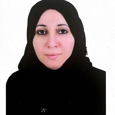
Rasha Hatem Saeed Dosh
Assistant Professor

Rasha Hatem Saeed Dosh

0Publications
0Lectures
0Interests

About

I obtained a bachelor degree from the University of Babylon / College of Science / department of biology in 1994. I worked as a biologist at the University of Kufa / College of Medicine / department of Anatomy and Histology. I received the title of assistant lecturer in the college of medicine after obtaining a master's degree from Al-Mustansiriya University / college of science / department of biology in 2000. As a result of my research work, I became a lecturer in 2007.
In 2014, I obtained a grant from the Ministry of Higher Education and Scientific Research in Iraq to complete my postgraduate studies outside the country in the United Kingdom, where I enrolled for a PhD in the field of tissue engineering and biotechnology of stem cell at Sheffield Hallam university / Department of biomolecular sciences and chemistry and obtained a PhD in 2018.
During my study in the UK, I acquired a lot of skills and experience in using the latest equipment and doing the latest biological tests in my field of study. I was selected to train postgraduate and undergraduate students in the department, in addition to gaining valuable experience in academic research work, and I published four research papers and an academic article in valuable international journals. In addition, I participated in many international conferences to present the results of my research as oral presentations in Canada, Spain and the UK, and in the form of posters in Canada, Spain, the UK and Switzerland. I was awarded the third best poster award at a conference in the UK, and one of the pictures of my academic articles was chosen as the cover for the journal that published this article. I was awarded three letters of appreciation and an award by the President of the University of Kufa for my researches which were published in Scopus first quarter journal

Publications Since 2014
Review Paper
1- Dosh R. H., Jordan-Mahy N., Sammon C., and Le Maitre C. L. Tissue engineering laboratory models of the small intestine. Tissue Engineering Part B. 2018, 24(2), 98-111.
DOI: 10.1089/ten.teb.2017.0276
A schematic diagram illustrating the morphology of the small intestinal layers was chosen as the art cover for the journal.
2- Research Papers
1- • Dosh R. H., Essa A., Jordan-Mahy N., Sammon C., and Le Maitre C. L. Use of hydrogel scaffolds to develop an in vitro 3D culture model of human intestinal epithelium. Acta Biomaterilia, 2017, 62, 128-143.
http://dx.doi.org/10.1016/j.actbio.2017.08.035
2- Dosh R. H., Jordan-Mahy N., Sammon C., and Le Maitre C. L. Long-term in vitro 3D hydrogel co-culture model of inflammatory bowel disease. Scientific Reports. 2019, 9:1812. https://doi.org/10.1038/s41598-019-38524-8
3- Dosh R. H., Jordan-Mahy N., Sammon C., and Le Maitre C. L. Interleukin-1 is a key driver of inflammatory bowel disease - demonstration in a murine IL-1Ra knockout model. Oncotarget, 2019,
Vol. 10, (No. 37), 3559-3575
• DOI: 10.18632/oncotarget.26894
4- Dosh R. H., Jordan-Mahy N., Sammon C., and Le Maitre C. L. Use of L-pNIPAM hydrogel as a 3D-scaffold for intestinal crypts and stem cell tissue engineering. Biomaterials Science. 2019, 7, 4310.
DOI: 10.1039/c9bm00541b
Published abstracts
• Dosh R. H., Jordan-Mahy N., Sammon C., and Le Maitre C. L. (2017)
The use of pNIPAM-laponite® hydrogel in 3D-cell culture of the intestine.
European Cells and Materials. Abstract, 33 Suppl. 2 (P517).
Published abstracts
• Dosh R. H., Jordan-Mahy N., Sammon C., and Le Maitre C. L. (2017)
The use of pNIPAM-laponite® hydrogel in 3D-cell culture of the intestine.
European Cells and Materials. Abstract, 33 Suppl. 2 (P517).
Conferences
Oral Presentations
1-January 2017 - Royal Society of Chemistry Biomaterials, Belfast, UK: Novel in vitro three-dimensional culture of human intestinal epithelium. Dosh R. H., Jordan-Mahy N., Sammon C., and Le Maitre C. L.

2-October 2016 - 6th Clinical Gastroenterology and Hepatology, Toronto, Canada: Novel in vitro three-dimensional culture of human intestinal epithelium. Dosh R. H., Jordan-Mahy N., Sammon C., and Le Maitre C. L.

3-October 2016 - 9th Euro - Global Gastroenterology Conference, Valencia, Spain: Novel in vitro three-dimensional culture of human intestinal epithelium. Dosh R.H., Jordan-Mahy N., Sammon C., and Le Maitre C. L.
Poster Presentations
1- September 2017 - 1st Iraqi Society Conference, Sheffield. UK: The use of pNIPAM-laponite hydrogel in 3D cell culture of the intestine. Dosh R.H., Jordan-Mahy N., Sammon C., and Le Maitre C. L. (3rd Place Poster Prize).

2-July 2017 - TCES, Manchester, UK: Development of a 3D in vitro co-culture cells model for studying inflammatory bowel disease. Dosh R.H., Jordan-Mahy N., Sammon C., and Le Maitre C. L.

3-June 2017 - TERMIS, Davos, Switzerland: The use of pNIPAM-laponite hydrogel in 3D cell culture of the intestine. Dosh R.H., Jordan-Mahy N., Sammon C., and Le Maitre C. L.

4-December 2016 - BMRC Winter Poster Session, Sheffield, UK: Interleukin-1 receptor antagonist knockout mice as a model of the inflammatory bowel disease. Dosh R.H., Jordan-Mahy N., Sammon C., and Le Maitre C. L.

5-October 2016 - 6th Clinical Gastroenterology and Hepatology, Toronto, Canada: Interleukin-1 receptor antagonist knockout mice as a model of the inflammatory bowel disease. Dosh R.H., Jordan-Mahy N., Sammon C., and Le Maitre C. L.

6-October 2016 - 9th Euro-Global Gastroenterology Conference, Valencia, Spain: Interleukin-1 receptor antagonist knockout mice as a model of the inflammatory bowel disease. Dosh R.H., Jordan-Mahy N., Sammon C., and Le Maitre C. L.

7-July 2016 - Health and Wellbeing Faculty Research Day, Sheffield, UK: Novel in vitro three-dimensional culture of human intestinal epithelium. Dosh R.H., Jordan-Mahy N., Sammon C., and Le Maitre C. L.

8-January 2016 - Royal Society of Chemistry Biomaterials, Birmingham, UK: In vitro three-dimensional hydrogel culture of human intestinal cell lines to develop a 3D model. Dosh R.H., Jordan-Mahy N., Sammon C., and Le Maitre C. L.
9-December 2015 - BMRC Winter Poster Session, Sheffield, UK: In vitro three-dimensional hydrogel culture of human intestinal cell lines to develop a 3D model. Dosh R.H., Jordan-Mahy N., Sammon C, and Le Maitre C. L.
Kufa Medical Virtual Medical Conference: May 31th, 2020

Research Interests

1

Publications

15
2026

Anti-inflammatory and biocompatibility effects of Portulaca oleracea and nanoparticles on the bone

AuthorsRasha Hatem Dosh, Najat Mutar Alsallami, Manar Mudhafar Alnema, Siham Mahmood Al-Rehemi, Bushra Habeeb Al-Maula
JournalNatural Resources for Human Health
Date2026-08-27
Abstract

Background: Bone healing depends on a well-balanced and highly coordinated process of inflammation, repair, and bone remodeling. Portulaca oleracea and gold nanoparticles (Au-NPs) have demonstrated potential applications in wound healing in soft tissues; however, their potential in hard-tissue bone regeneration has yet to be studied in comparison with each other and within the context of biocompatibility. Objective: to assess and compare the anti-inflammatory and biocompatibility effects of Portulaca oleracea extract and Au-NPs on early bone healing in a standardized defect model of tibia of the rabbit. Materials and Methods: Cortical bone defects were surgically created in the tibiae of rabbits and assigned to three independent groups: topical Portulaca oleracea extract treatment, Au-NP treatment, and untreated control. Histological and histomorphometric assessments were conducted at critical early time points (days 3 and 7 post-surgery) to determine the inflammatory cell dynamics, proliferation of fibroblasts, osteoblast and osteocyte activity, and neovascularization. Results: Morphometric analysis of the bone defect area showed that the application of Portulaca oleracea extract and gold nanoparticles (Au-NPs) had a significant effect on the process of tissue healing and osteogenesis. All experimental groups had a significant decrease in the number of inflammatory cells and a significant increase in blood vessel density and in the number of fibroblasts by day 7, when compared with the control group. Importantly, the treatment with Portulaca oleracea achieved the highest vascularization at day 3, and reduced inflammation significantly at day 7. In addition, both interventions resulted in early recruitment of osteoblasts at day 3 and Au-NP treatment resulted in the highest osteocyte density at day 7. Conclusion: Results suggest that the bone defect repair process is promoted by the extract of Portulaca oleracea and Au-NPs in independent but synergic cellular mechanisms during early stages. Both Au-NPs and Portulaca oleracea are highly biocompatible and are potential therapeutic agents for bone tissue regeneration.

2026

Effects of gold nanoparticles and apricot seeds on bone healing in New Zealand rabbits

AuthorsZena J. Wally, Bushra H. Al-Maula, Sahar G. AL-Kazzaz, Rasha H. Dosh, Wasna’a M. Abdulridh
JournalJournal of Taibah University Medical Sciences
Date2026-01-13
2026

Enhanced wound healing in rat cheeks treated with lavender and ginger oils: A histological study

AuthorsEnas F. Kadim, Zena Wally, Rasha Dosh, Julfikar Haider
JournalJournal of Taibah University Medical Sciences
Date2026-03-12
2024

Histopathological, immunohistochemical and physiological study for the hepatoprotective effect of melatonin against inhrifampicin-induced hepatotoxicity in mice model

AuthorsSiham Mahmood Al-Rehemi, Rasha Hatem Dosh, Maha Jamal Frayyeh, Rihab Hameed Al Mudhafar, Hussein Abdulkadhim
JournalWiadomości Lekarskie Medical Advances
Date2024-08-12
Abstract

Aim: The purpose of this study is to assess the hepatoprotective effect of melatonin against isoniazid (INH) and rifampicin (RMP) induced hepatotoxicity in albino mice. Materials and Methods: Adult male mice were divided into four groups: saline, INH-RMP, INH-RMP+MT and MT were administered for 21 days. Biochemical analyses were performed for the determination of ALT, AST. Histopathological changes in the liver and Immunohistochemical assessment to determine the expression of Caspace3 were also examined. Results: Biochemical analysis revealed significant increases in serum ALT and AST in INH-RMP group. Histopathological findings demonstrated severe liver damage in INH-RMP group as compared with control group. In contrast, treatment of mice with melatonin (MT) markedly mitigated the liver injury. Immunohistochemical findings demonstrated apoptotic marker caspace3 significantly higher in INH-RMP group as compared with control group. Conclusions: Experimental findings highlight the potential benefits of melatonin in this model, prompting speculation on its potential application in human therapy.

2023

Histologicalevaluationof theeffectsofbonemorphogeneticprotein9 andangiopoietin1onbonehealing

AuthorsEnasFadhil, Rasha H.Dosh, Zena J.Wally, Julfikar Haider
JournalJournal of Taibah University Medical Sciences
Date2023-02-16
Abstract

Objectives: Bone healing remains a critical clinical or thopedicproblem.Bone,whichisagreatlyvascularized tissue, depends on the tight temporal and spatial link betweenbloodvesselsandbonecells.Thus,angiogenesis iscrucial forskeletalgrowthandbonefracturehealing. Thepurposeofthisstudywastoevaluatetheefficacyof thelocalapplicationofosteogenicandangiogenicfactors such as bone morphogenetic protein 9 (BMP9) and angiopoietin1 (Ang1), respectively, andtheir combina tionasanosteoinducer intheprocessofbonehealing. Methods: Forty-eight male albino rats, weighing 300 e400gandaged6e8months,wereutilizedinthisstudy. Theanimalsunderwentsurgeryonthemedialsideofthe tibiabone. In the control group, anabsorbablehemo staticspongewaslocallyappliedtothebonedefect,while experimentalgroupswereseparatedintothreegroups.In GroupI,1mgBMP9was locallyapplied,GroupIIwas treatedwith1mgAng1,andGroupIIIwastreatedwith local application of a combination (0.5 mg BMP9 and 0.5 mg Ang1). All experimental groups were fixed with an absorbable hemostatic sponge. The rats were sacrificed on days 14 and 28 after surgery. Results: Local application of BMP9 alone, Ang1 alone, and their combination to a tibia defect caused osteoid tissue formation and significantly increased the number of bone cells. A gradual decrease in the number of trabecular bone, an increase in trabecular area, and no significant difference in the bone marrow area were noted. Conclusion: The combination of BMP9 and Ang1 has therapeutic potential in promoting the healing process of bone defects. Osteogenesis and angiogenesis are regulated by BMP9 and Ang1. These factors act together to accelerate bone regeneration more efficiently than either factor alone

2022

IN VIVO ACTIVATION OF P2Y4 PURINERGIC RECEPTORS USING ATP IN RAT EPIDERMAL TISSUE

AuthorsRasha Hatem Dosh, Hydar Muhsin Khalfa, Siham Mahmood Al-Rehemi, Hayder Lateef Almsaid,
JournalWiadomości Lekarskie
Date2022-11-04
Abstract

The aim: This study was carried out to examine the presence of P2Y4 receptors in rat epidermal tissue and how their in vivo activation leads to histological and genetic changes. Materials and methods: Thirty-six Wistar rats were separated into six groups each of six rats, the control group and ve injected groups with increasing concentrations of ATP intradermally (0.1, 5.0, 10.0, 50.0, 100.0 µg/ml). The histological and genetic examination was performed from excised tissues. Results: Noticeable histological thickening of the epidermal layer in rats injected with high concentrations of ATP. No apparent histological damage was seen in all injected groups. The genetic expression seems to also increase following exposure to variable concentrations of ATP. Conclusions: Purinergic receptors activated by ATP molecules are highly involved in the development of adult tissues. Their precise location within the epidermal layer indicated their importance in cellular proliferation and di erentiation of epidermal cells. Excessive exposure to ATP results in their robust genetic ectopic over expression indicative of increased cellular activity.

2021

Assessing the Effects of Administering Calcium Oxide Nanoparticles on Oral Mucosa and Tongue of Male Wister Rats

AuthorsBushra Habeeb Al-Maula, Zena Wally, Rasha Dosh, Abtesam Imhemed Aljdaimi
JournalNano Biomed Eng
Date2021-09-09
Abstract

The present study was conducted to evaluate the impact of calcium oxide-nanoparticles (CaO-NPs) administration at different concentrations on the epithelial thickness, papilla length and number of blood vessels of male Wister rats’ buccal mucosa, and the epithelial thickness, filiform length and number of blood vessels of the rats’ tongue. Twelve rats were used and randomly allocated into four groups: control group (untreated) and the three experimental groups were orally treated with CaO NPs at different doses 25, 50 and 100 mg/kg of the body weight over two months. On the 61st day, all the animals were sacrificed, and 1 cm of the buccal mucosa and the body of the tongue were carefully removed for histological analysis. Histological tissues were studied under a light microscope to investigate the impact of CaO-NPs administration on the oral tissues. An image processing software (Image J) was used to measure the epithelial thickness, papilla length and number of blood vessels of the rats’ buccal mucosa, and the epithelial thickness, filiform length and number of blood vessels of the rats’ tongue. The results showed that the CaO nanoparticles administration caused epithelial atrophy and decreased vascularization of buccal and tongue mucosa with all tested doses of CaO-NPs and the 100 mg/kg concentration showed the most significant effect.

2021

Studying Effects of Calcium Oxide Nanoparticles on Dentinogenesis in Male Wistar Rats

AuthorsBushraHabeebAl-Maula, Mohanad Jameel Najm Al-Magsoosi, Zena Jehad Wally, Rasha Hatem Dosh
JournalInternational Journal of Dentistry
Date2021-07-26
Abstract

/is study aimed to evaluate potential impacts of calcium oxide nanoparticles (CaO-NPs) at different dosages on predentin thickness, number of blood vessels, periodontal ligament thickness, and blood glucose level of Wistar rats. Twelve rats were randomly gathered into four groups, untreated (control) and CaO-NP-treated groups at three concentrations (25, 50, and 100mg/ kg of the body weight) over a period of 60 days. Histological investigation was performed on twenty-four lower incisor teeth extracted from all the tested groups under a light microscope, and an automatic Fujifilm was used to measure the blood glucose level. /e results showed that regular nanoparticle treatment significantly increased predentin and periodontal ligament thicknesses, a gradual decrease in vascularization in the pulp tissue, and an increase in the blood glucose level as the dosages of nanoparticles administered to the rats increased. Administration of the CaO-NPs at low dosage (25mg/kg) could be beneficial for the growth and integrity of teeth and dentinal tissues in rats

2021

Association of Toll-like receptors 4 (TLR-4) gene expression and polymorphisms in patients with severe asthma

AuthorsWasan Shukur , Kifah Alyaqubi, Rasha Dosh, Ali Al-Ameri, Hayder Al-Aubaidy
JournalJOURNAL of MEDICINE and LIFE
Date2021-07-30
Abstract

Innate immunity plays a central role in the pathogenesis of severe asthma, and it is closely linked to elevated IgE and Toll-like receptor 4 (TLR-4) levels. However, there is a scarcity of information about the association of the TLR-4 receptor polymorphism in the pathogenesis of severe asthma. This study highlights the level of gene expres sion of different alleles in asthmatic patients compared to healthy control individuals. This was a randomized control trial, which included 150 patients with asthma (with high serum levels of IgE) with a matching 150 healthy control individuals. Partici pants had a series of blood tests to measure various immune parameters: interleukin-1 (IL-1), interleukin-6 (IL-6), tumor necrosis factor (TNF), intercellular adhesion mole cule-1 (ICAM1) and detect allele type and gene expression of the TLR-4 gene. Patients with asthma had significantly higher levels of IL-8 when compared to the healthy con trol participants. In addition, in the rs91 genotyping, there were significant differences in the levels of IL-8 and TNF between CC and TT genotyping. While in rs90 TLR-4, TNF levels were significantly higher in AA vs. AG and GG genotypes among the asth matic patients when compared to the control group. The results showed that in TLR-4, rs4986791 were significantly associated with asthma risk. Polymorphisms in TLRs play essential roles in asthma.

2021

Gene expression of carbonic anhydrase 9 (CA9) in de novo acute leukemia as a predictive marker for prognosis

AuthorsKifah Jabbar Alyaqubi, Rasha Hatem Dosh, Rawaa Behlul Al-Fatlawi, Siham Mahmood Al-Rehemi
JournalJOURNAL of MEDICINE and LIFE
Date2021-08-07
Abstract

Carbonic anhydrase 9 (CA9) is a marker for decreased O2 concentration and acidosis, associated with poor prognosis in cancerous patients. The current study suggested that the changes in CA9 gene expression level might be used as a predictive marker to assess early prognosis at the time of detection of de novo leukemia, and then monitor tumor prog ress during treatment. This study highlights the level of CA9 gene expression in leukemic patients. A total of 44 cases (acute myeloid leukaemia (AML) group: 23 cases; acute lymphoid leukaemia (ALL) group: 13 cases; control group: 8 healthy volunteers) were selected for this study. The CA9 gene expression was assessed by a real-time PCR with the SYBR green assay. A high level of CA9 gene expression was noticed in AML patients compared to the control group, while the results were not significant in ALL patients. After treatment follow-up, significant differences were observed in CA9 gene expression between a complete response and no response in AML patients. As a result, the CA9 tumor gene could act as a potential early marker for acute leukemia prognosis. A low level of CA9 expression was associated with better clinical outcomes, while a high level was related to a negative prognosis in patients with AML

2019

Use of L-pNIPAM hydrogel as a 3D-scaffold forintestinal crypts and stem cell tissue engineering

AuthorsRasha Dosh, Nicola Jordan-MahyChris Sammon Christine Le Maitre
JournalBiomaterials science
Date2019-01-08
Abstract

Intestinal stem cells hold great potential in tissue regeneration of the intestine, however, there are key limitations in their culture in vitro. We previously reported a novel synthetic non-biodegradable hydrogel as a 3D culture model for intestinal epithelium using Caco2 and HT29-MTX cells. Here, we investigated the potential of this system as a 3D scaffold for crypts and single intestinal stem cells to support long-term culture and differentiation. Intestinal crypts were extracted from murine small intestines and Lgr5+ stem cells isolated by magnetic activated cell sorting. Crypts and stem cells were suspended within Matrigel or L-pNIPAM for 14 days or suspended within Matrigel for 7 days then released, dissociated, and suspended within, or on L-pNIPAM hydrogel for 28 days. Cellular behaviour and phenotype were determined by histology and immunohistochemistry for stem cell and differentiation markers: Lgr5, E-cadherin MUC2 chromograninA and lysozymes. Isolated crypts and Lgr5+ intestinal stem cells formed enteroids with a central lumen surrounded by multiple crypt-like buds when cultured in Matrigel. In contrast, when crypts and stem cells were directly suspended within, or layered on L-pNIPAM hydrogel under dynamic culture conditions they formed spherical balls of cells, with no central lumen. When enteroids were initially formed in Matrigel from crypts or single Lgr5+ intestinal stem cells and dissociated into small fragments or single cells and transferred to L-pNIPAM hydrogel they formed new larger enteroids with numerous crypt-like buds. These crypt-like buds showed the presence of mucin-producing cells, which resembled goblet cells, scattered throughout their structures. Immunohistochemistry staining also showed the expression of Lgr5 and differentiation markers of all the main intestinal cell types including: enterocytes, goblet cells, enteroendocrine and Paneth cells. This demonstrated that L-pNIPAM hydrogel supported long-term culture of crypts and Lgr5+ stem cells and promoted intestinal cell differentiation.

2019

Long-term in vitro 3D hydrogel co-culture model of inflammatory bowel disease

AuthorsRasha Dosh, N, JordaN-MahyC, SammonC, Le Maitre
JournalScientific Reports
Date2019-02-12
Abstract

The in vitro study of the pathogenesis of inflammatory bowel disease (IBD) requires a cell model which closely reflects the characteristics of the in vivo intestinal epithelium. This study aimed to investigate the application of L-pNIPAM hydrogel as a scaffold to develop a long-term 3D co-culture model of Caco-2 and HT29-MTX cells under conditions analogous to inflammation, to determine its potential use in studying IBD. Monocultures and co-cultureswere layered on L-pNIPAM hydrogel scaffolds and maintained under dynamic culture conditions for up to 12 weeks. Treatments with IL-1β, TNFα, and hypoxia for 1 week were used to create an inflammatory environment. Following prolonged culture, the metabolic activity of Caco-2 monoculture and 90% Caco-2/10% HT29-MTX co-cultures on L-pNIPAM hydrogels were increased, and finger-like structures, similar in appearance to villi were observed.Following treatment with IL-1β, TNFα and hypoxia, ALP and ZO-1 were decreased, MUC2 increased, and MUC5AC remained unchanged. ADAMTS1 was increased in response to hypoxia. Caspase 3 expression was increased in response to TNFα and hypoxic conditions. In conclusion, L-pNIPAM hydrogel supported long-term co-culture within a 3D model. Furthermore, stimulation with factors seen during inflammation recapitulated features seen during IBD.

2019

Interleukin 1 is a key driver of inflammatory bowel disease-demonstration in a murine IL-1Ra knockout model

AuthorsRasha Dosh, Nicola Jordan-MahyChris SammonChristine Le Maitre
Journaloncotarget
Date2019-05-28
Abstract

Interleukin 1 (IL-1) is an important mediator of inflammation and tissue damage in inflammatory bowel disease (IBD). The balance between IL-1 and IL-1Ra as a natural inhibitor plays a vital role in a variety of diseases. Here, we investigated whether changes seen during IBD are induced spontaneously in mice lacking a functional IL1rn gene. Histological staining was performed on the jejunum and ileum of BALB/c IL-1rn+/+ and IL-1rn-/- mice to characterize crypt-villus height, villus width, and number of goblet cells per villus. Pro-inflammatory cytokines, immune cell infiltration and matrix-degrading enzymes, together with the production of intestinal enzymes and the integrity of tight and adherent junction proteins were determined using immunohistochemistry. In the small intestine of BALB/c IL-1rn-/- mice the villus heights were significantly reduced; and in the ileum this was accompanied by a decrease in villi width. There was also an increase in goblet cell number and mucin production compared to wild-type mice. IL-1α and IL-1β immunopositivity were increased, whilst IL-1R1 expression was decreased in IL-1rn-/- mice. IL-15 and TNFα were also increased in older IL-1rn-/- mice. Increased polymorphonuclear and macrophage infiltration were seen in IL-1rn-/- mice, whilst expression of matrix-degrading enzymes and digestive enzymes were unchanged, except for dipeptidyl peptidase IV which was increased in younger IL-1rn-/- mice compared to wild type mice. The expression of tight and adhesion junctions were also dramatically decreased in IL-1rn-/- mice. In conclusion, IL1rn-/- mice developed spontaneous abnormalities which displayed features associated with IBD, demonstrating a clear role for IL-1 in IBD.

2017

Tissue Engineering Laboratory Models of the Small Intestine

AuthorsRasha Dosh, Nicola Jordan-MahyChris SammonChristine Le Maitre
JournalTissue Eng Part B Rev
Date2017-11-27
Abstract

In recent years, three-dimensional (3D) cell culture models of the small intestine have gained much attention. These models support cell proliferation, migration, and differentiation and encourage tissue organization which is not possible in two-dimensional (2D) culture systems. Furthermore, the use of a wide variety of cell culture scaffolds and support substrates has revealed considerable differences in cell behavior and tissue organization. These systems have been used in combination with intestinal stem cells, organoid units, or human colonic adenocarcinoma cell lines such as Caco-2 and HT29-MTX to generate a number of in vitro and in vivo models of the intestine. In this study, we review the current 2D and 3D tissue engineering models of the intestine to determine the most effective sources of intestinal cells and current research on support scaffolds capable of inducing the morphological architecture and function of the intestinal mucosa.

2017

Use of hydrogel scaffolds to develop an in vitro 3D culture model of human intestinal epithelium

AuthorsRasha Dosh, A, EssaN, Jordan-MahyC, SammonC, Le Maitre
JournalActa Biomaterialia
Date2017-08-30
Abstract

The human intestinal cell lines: Caco-2 and HT29-MTX cells have been used extensively in 2D and 3D cell cultures as simple models of the small intestinal epithelium in vitro. This study aimed to investigate the potential of three hydrogel scaffolds to support the 3D culture of Caco-2 and HT29-MTX cells and critically assess their use as scaffolds to stimulate villi formation to model a small intestinal epithelium in vitro. Here, alginate, l-pNIPAM, and l-pNIPAM-co-DMAc hydrogels were investigated. The cells were suspended within or layered on these hydrogels and maintained under static or dynamic culture conditions for up to 21 days. Caco-2 cell viability was increased when layered on the synthetic hydrogel scaffolds, but reduced when suspended within the synthetic hydrogels. In contrast, HT29-MTX cells remained viable when suspended within or layered on all 3D cultures. Interestingly, cells cultured in and on the alginate hydrogel scaffolds formed multilayer spheroid structures, whilst the cells layered on synthetic hydrogels formed villus-like structures. Immunohistochemistry staining demonstrated positive expression of enterocyte differentiation markers and goblet cell marker. In conclusion, l-pNIPAM hydrogel scaffolds supported both cell lines and induced formation of villus-like structures when cells were layered on and cultured under dynamic conditions. The ability of the l-pNIPAM to recapitulate the 3D structure and differentiate main cell types of human intestinal villi may deliver a potential alternative in vitro model for studying intestinal disease and for drug testing.

Lectures

5

Head and Neck

MSK

Histology of GIT

TOB

Epithelial Tissue

Histology

Histology

View details